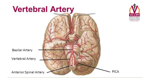

In addition, on the right side one PICA coming out from the hypoplastic VA and one AICA coming out from the BA (Figure 1). The BA was viewed as a continuation of the dominant left VA (Figure 1). The external diameters of the left VA were approximately 5 mm, the diameter of the right one was measured approximately 2 mm. The diameters of the two intracranial segments of VA’s were measured. Results and Case Reportĭuring routine dissection of head of 75-year-old male cadaver we observed the intracranial segment of right VA diameter was thinner (hypoplastic) than the intracranial segment of left VA (dominant). The sections were stained with Masson trichrome and histological examination was performed under a light microscope. 5 µm sections were obtained using a microtome. They were fixed in 10% formalin and embedded in paraffin. We believe that the findings of the present case of aberrant vessels may influence the surgical procedures and better appreciate the arterial flow of the right and the left-brain in clinical care. In the present case, at the vertebrobasilar junction a dominant and hypoplastic VA and, duplicate PICA and AICA was observed at the same time in one cadaver. There are studies that show decreased diameter of the VA is closely related to some clinical features such as vertigo, migraine, and tinnitus. Clinically, it has been shown that, any presence of occlusion, stenosis, or low amount of blood flow in the VA, BA or its branches may lead to the vertebrobasilar arterial insufficiency. The vertebrobasilar system is composed of bilateral vertebral arteries (VA), unpaired basilar artery (BA) and its branches, which, supplies the cervical spinal cord, brainstem, cerebellum, thalamus, and occipital lobes. Keywords: Anatomic variation Vertebral artery Basilar artery Posterior inferior cerebellar artery Anterior inferior cerebellar artery This is the first report concerning the dominant and hypoplastic VA and duplicate PICA and AICA at the same time in one cadaver. Understanding the variation of the arterial vertebrobasilar system may influence the surgical procedures and better appreciate the arterial flow of the right and the left-brain in clinical care. The external diameters of the left VA were over 2 times more than the right one. We examined the histological sections of the dominant and hypoplastic VA. During routine dissection in the department of Anatomy, the presence of a very thin hypoplastic right VA, the left dominant VA was continuous with the basilar artery, and both duplicate of left PICA and left AICA were observed.

Sections were stained with Masson’s trichrome and histological examination was performed under the light microscope. Artery sections of 5 µm were obtained for morphological and microscopic examinations. To describe aberrant dominant and hypoplastic vertebral (VA) arteries and duplicated posterior inferior cerebellar artery (PICA) and anterior inferior cerebellar artery (AICA).


 0 kommentar(er)
